Quantcast
Channel: cancer Archives - VAI
Viewing all 60 articles
Browse latest View live

Van Andel Institute scientists model complex cancer cell behavior

$
0
0

Cell cannibalism process implicated in human disease determined through collaboration with Los Alamos National Laboratory

Grand Rapids, Mich. (March 14, 2013) – Van Andel Institute (VAI) researchers recently published a mathematical model of autophagy, a complex process involved in determining cell death or survival.  The autophagic process has been implicated in human diseases such as neurodegeneration, and has recently become a dynamic topic in the field of cancer research.

Autophagy is sometimes referred to as cannibalism within the cell and is a Greek term that literally translates as “self-eating.”  This complex cellular behavior generates a multitude of questions and challenges for researchers.  Jeffrey P. MacKeigan, Ph.D., senior author of the study, recognized that understanding cellular events requires more than basic biology experiments and embarked on a collaboration with computational scientists at Los Alamos National Laboratory (LANL).

“By joining forces and taking a multidisciplinary approach, we aim to more rapidly advance our knowledge of autophagy and translate those findings into a positive impact on human health,” said MacKeigan, Head of VAI’s Laboratory of Systems Biology.

The mathematical model, published by the collaborative team in the journal Autophagy, uses an adaptation of the Monte Carlo method, a type of computational algorithm relying on random sampling that Los Alamos scientists used after World War II to assist with the complexities of nuclear fission devices like the atomic bomb.   In partnership with William Hlavacek, Ph.D., at LANL, MacKeigan’s team used the model to make accurate predictions about autophagy in cancer cells.

“These studies mark the first steps toward a more robust and comprehensive model,” MacKeigan said. “We hope to further cultivate the model in order to inform future treatment strategies, and are working to secure funding to make these enhancements.”

The role of autophagy in cancer and other human diseases is yet to be completely understood.  To tackle this and other biological challenges, MacKeigan and his lab use a “systems biology” approach.  By leveraging an arsenal of cutting-edge methods and collaborating with experts in complementary fields, these researchers are working to make significant contributions to the study of autophagy.

Katie Martin, Ph.D., lead author of the study and scientific project leader in VAI’s Laboratory of Systems Biology, explains the research in the context of human health:

“Cancer cells can often adapt to survive chemotherapy,” Martin said. “If our model can predict how autophagy contributes to this survival, we could provide valuable information for the design of more effective therapies – and that’s exciting!”

###

Van Andel Institute

Established by Jay and Betty Van Andel in 1996, Van Andel Institute (VAI) is an independent research and educational organization based in Grand Rapids, Mich., dedicated to preserving, enhancing and expanding the frontiers of medical science, and to achieving excellence in education by probing fundamental issues of education and the learning process.  Van Andel Research Institute (VARI), VAI’s research arm, is dedicated to studying the genetic, cellular and molecular origins of cancer, Parkinson’s and other diseases and working to translate those findings into effective therapies. This is accomplished through the work of more than 200 researchers in on-site laboratories and in collaborative partnerships that span the globe. Find out more about Van Andel Institute or donate by visiting www.vai.org.

The post Van Andel Institute scientists model complex cancer cell behavior appeared first on VAI.


Van Andel Institute cancer research study discovers a new strategy to target cancer cells

$
0
0

Grand Rapids, Mich. (Nov. 15, 2013) – A discovery by scientists at Van Andel Institute offers promise of an innovative treatment strategy to impair the growth of cancer cells. The study identifies two compounds that slowed tumor growth while protecting normal tissue. The results may lead to an improved and safer therapy for a variety of human cancers.

The new report, “Small molecule intramimics of formin auto-inhibition: a new strategy to target the cytoskeletal remodeling machinery in cancer cells,” was carried out at the Van Andel Research Institute in collaboration with scientists from Grand Valley State University and Kalamazoo Valley Community College’s Michigan High Throughput Screening Center and published this week in the journal Cancer Research.

“This discovery could lead to novel cancer therapies for hard to treat cancers and potentially serve as an alternative or an adjuvant to Taxol or Vinblastine, agents commonly used in chemotherapy to treat breast, ovarian, lung, testicular and certain blood cancers,” said Dr. Arthur S. Alberts, Ph.D., Professor and head of the Laboratory of Cell Structure and Signal Integration at Van Andel Institute and the senior author of the study.

Results
All cells have an internal structural framework that makes it possible for the cell to move and divide. This “cytoskeleton” is a valid target for currently used chemotherapeutic drugs like Taxol and Vinblastine. These drugs successfully target the building blocks of the cytoskeleton to keep cancerous cells from growing and dividing, which can prevent tumor growth.

The study describes a new class of compounds called Intramimics that target a family of proteins in the cell called formins. “Formins are the masons of the cell that assemble the individual building blocks into the structures that comprise the cytoskeleton,” explained Dr. Alberts, who has spent the past fifteen years studying the genetics, molecular and cell biology of formins in cancer and other diseases.

Because their mechanism of action is distinct from currently available chemotherapeutic agents, it is hoped that the Intramimic compounds can specifically target cancer cells and spare healthy cells without the dose-limiting side effects experienced with Taxol and Vinblastine.

The two Intramimic compounds identified in this study were shown to trigger stabilization of microfilaments and microtubules that make up the cellular cytoskeleton. Experiments on cancer cells showed that Intramimics affected changes in gene expression that are associated with impaired cell growth and programmed cell death (apoptosis), that would be expected to reduce tumor size or slow tumor growth. Indeed, in another experimental system, the Intramimics did slow tumor growth. Taken together these studies suggest this strategy will be effective for treating solid tumors. Other preliminary evidence suggests potential application in the treatment of blood cancers as well.

“This discovery provides a new development regarding clinically validated drug targets in an area of research where few new strategies have emerged,” Dr. Alberts said. “Intramimics will serve as leads for further exploration and pharmacological development.”

The complete paper can be found on the Cancer Research website.

###

About Van Andel Institute

Established by Jay and Betty Van Andel in 1996, Van Andel Institute (VAI) is an independent research and educational organization based in Grand Rapids, Mich., dedicated to preserving, enhancing and expanding the frontiers of medical science, and to achieving excellence in education by probing fundamental issues of education and the learning process.  Van Andel Research Institute (VARI), VAI’s research arm, is dedicated to studying the genetic, cellular and molecular origins of cancer, Parkinson’s and other diseases and working to translate those findings into effective therapies. This is accomplished through the work of more than 200 researchers in on-site laboratories and in collaborative partnerships that span the globe. Find out more about Van Andel Institute or donate by visiting www.vai.org

The post Van Andel Institute cancer research study discovers a new strategy to target cancer cells appeared first on VAI.

Researchers thwart cancer cells by triggering ‘virus alert’

$
0
0

Note: This press release was written by John Hopkins Medicine. Dr. Stephen Baylin, who holds a joint appointment at Van Andel Research Institute (VARI) and at Johns Hopkins University’s Sidney Kimmel Comprehensive Cancer Center, is the corresponding author on the paper referenced. Read the release.


(August 27, 2015) – Working with human cancer cell lines and mice, researchers at the Johns Hopkins Kimmel Cancer Center and elsewhere have found a way to trigger a type of immune system “virus alert” that may one day boost cancer patients’ response to immunotherapy drugs. An increasingly promising focus of cancer research, the drugs are designed to disarm cancer cells’ ability to avoid detection and destruction by the immune system.

In a report on the work published in the Aug. 27 issue of Cell, the Johns Hopkins-led research team says it has found a core group of genes related to both a viral defense warning system and susceptibility to a demethylating drug called 5-azacytidine that chemically alters their ability to operate through a process called demethylation.

A study with similar findings authored by Daniel De Carvalho, Ph.D., at the Ontario Cancer Institute/Princess Margaret Hospital, and Peter Jones, Ph.D., D.Sc., director of research at the Van Andel Institute, focused on the ability of DNA demethylating agents to target colorectal cancer stem cells, is published in the same journal issue.

Tumors are known to co-opt cellular gene-silencing systems that add tiny chemicals called methyl groups to areas of genes, thereby turning off the affected gene function. Such “epigenetic” control normally occurs in many genes, including ones that contain DNA leftover from previous exposures to viruses. When epigenetic control of these genes is removed, the virus-laden gene sequences are activated and trigger an alert to immune system cells that a virus has invaded.

“A main barrier to immune therapy success has been the tumor’s ability to keep the immune system from functioning against the cancer,” says study leader Stephen Baylin, M.D., the Virginia and Daniel K. Ludwig Professor of Cancer Research at the Kimmel Cancer Center. “The immune cells are there, but like an unarmed army, they hang around and do nothing. However, certain epigenetic processes that silence such viral defense genes can be reversed in tumor cells with a demethylating drug, making immunotherapies work more effectively to kill cancer cells.”

For their new study, Baylin and his team worked with laboratory-grown cell lines from human ovarian, colon and skin cancer, and the team led by De Carvalho worked with colon cancer cells. In the cancer cell lines, both teams found that the viral defense pathway can be turned on when the cells were exposed to 5-azacytidine. Once the pathway is activated, Baylin adds, the tumor cells release signaling proteins called interferons that rouse other cancer-fighting cells in the immune system.

Then, the Johns Hopkins team created a gene signature of the viral defense pathway. In tumor samples available from the National Cancer Institute’s Cancer Genome Atlas project, the scientists used the gene signature to distinguish between tumor samples with high expression of the pathway from those with low expression. Those with high expression may respond to certain immunotherapy drugs without the aid of 5-azacytidine, but those with low expression levels may need the epigenetic drug to boost response to immunotherapy, says Baylin.

Looking for the connection between the pathway’s expression and immunotherapy drug response, the Johns Hopkins investigators and their colleagues focused on expression levels of the viral defense pathway in tumor cells from 21 patients with melanoma treated with the immune therapy drug ipilimumab at Memorial Sloan Kettering Cancer Center. They found high expression levels in the cells of seven of eight of those patients who had responded well to ipilimumab. Cells from all 12 patients with limited response to ipilimumab had low expression of the viral defense pathway.

In a melanoma mouse model in which ipilimumab alone was partially effective, adding 5-azacytidine to ipilimumab triggered a better tumor response.

“Our findings further decipher the mechanisms that lead to this tumor cell immune reaction and offer a way to potentially boost the success of immune therapies in patients with cancer,” says Baylin, who first became interested in the immune system’s connection to 5-azacytidine when laboratory research and clinical trials at Johns Hopkins hinted at the drug’s ability to prevent cancer cells’ proliferation when combined with immunotherapy.

Baylin and his colleagues say that, if their findings are confirmed and extended in clinical trials, the 5-azacytidine treatment could be followed by ipilimumab or other types of immunotherapy called checkpoint blockade, which lower cancer cells’ defenses and allow immune system cells to see and destroy them.

“Treatment with 5-azacytidine activates interferon signaling in tumor cells and, when followed by checkpoint blockade immune therapy, the immune cells could go into increased action against the cancer,” says Johns Hopkins research fellow and lead author Katherine Chiappinelli, Ph.D.

Baylin and Chiappinelli caution that clinical trials will take time to learn how effective the strategy of alerting the viral defense pathway might be. But the strategy holds promise, he says, for patients who have cancers with low expression of the pathway.

###

In addition to Baylin and Chiappinelli, other investigators included Pamela L. Strissel, Alexis Desrichard, Huili Li, Christine Henke, Benjamin Akman, Alexander Hein, Neal S. Rote, Leslie M. Cope, Alexandra Snyder, Vladimir Makarov, Sadna Buhu, Dennis Slamon, Jedd D. Wolchok, Drew M. Pardoll, Matthias W. Beckmann, Cynthia A. Zahnow, Taha Mergoub, Timothy A. Chan and Reiner Strick. In addition to the Johns Hopkins Kimmel Cancer Center, participating institutions included the University-Clinic Erlangen in Germany, Memorial Sloan Kettering Cancer Center, Case Western Reserve University and Jonsson Comprehensive Cancer Center at UCLA.

Baylin is a consultant for MDxHealth, which makes an assay procedure that is licensed to MDxHealth by The Johns Hopkins University. Baylin and the university are entitled to royalty shares from sales of the assay. Pardoll is a consultant for Pfizer and Amplimmune.

The research was funded by the National Institutes of Health’s National Cancer Institute (CA058184, F32CA183214), the Stand Up To Cancer Epigenetic Dream Team, the Hodson Trust, the Samuel Waxman Cancer Research Foundation, the Dr. Miriam and Sheldon G. Adelson Medical Research Foundation, a Department of Defense Teal Award (BC031272-SBB), the Pershing Square Sohn Cancer Research Alliance, the STARR Cancer Consortium, the Ludwig Foundation and German Cancer Aid.

Johns Hopkins Medicine
Media Relations and Public Affairs
Media contacts:
Patrick Smith, 410-955-8242, pjsmith@jhu.edu
Jania Matthews, 410-955-5384, jmatth27@jhmi.edu


The post Researchers thwart cancer cells by triggering ‘virus alert’ appeared first on VAI.

Researchers mimic viral infection in colon cancer stem cells, identify druggable target to potentially root out disease, end relapse

$
0
0

Note: This press release was written by Princess Margaret Cancer Centre. Dr. Peter Jones, Van Andel Research Institute’s (VARI) research director, is a contributing author on the study, which was led by Princess Margaret Cancer Centre and University of Toronto’s Dr. Daniel De Carvalho. Read the release.


(TORONTO, Canada – Aug. 27, 2015) – ​Researchers targeting colorectal cancer stem cells – the root cause of disease, resistance to treatment and relapse – have discovered a mechanism to mimic a virus and potentially trigger an immune response to fight the cancer like an infection.

The discovery, published online today in Cell, illuminates a major shift in understanding anti-tumour mechanisms and identifies a promising druggable target against colorectal cancer stem cells, says principal investigator and lead author Dr. Daniel De Carvalho, a scientist at Princess Margaret Cancer Centre, University Health Network. He is also Assistant Professor in the Department of Medical Biophysics, Faculty of Medicine at University of Toronto.

“By mimicking a virus the potential is to trick the immune system into ‘seeing’ the cancer cells as an infection that needs to be destroyed,” says Dr. De Carvalho. “Our work demonstrates that viral mimicry is a viable anti-tumour strategy.” Currently, colorectal ‘cancer recurs in about 50 per cent of patients and is among the top three leading causes of cancer-related deaths.

In the laboratory, the research team replicated human colorectal cancer in pre-clinical experiments and used bioinformatics analysis to demonstrate that a low-dose of the chemotherapy drug decitabine targeted the cancer stem cells by inducing viral mimicry.

Decitabine is approved by the U.S. Food and Drug Administration to treat myelodysplastic syndromes and leukemia, and for use in clinical trials for several types of solid-tumour cancers including colorectal. In Dr. De Carvalho’s research, the team discovered that this drug – known as an epigenetic therapy because it chemically modifies DNA – activates a pathway that recognizes viruses.

“We have found a switch to turn on an anti-viral response in colorectal cancer stem cells, which seem to be especially sensitive to it,” says Dr. De Carvalho. This discovery builds on earlier published research from other Princess Margaret scientists, Dr. John Dick, the pioneer of the cancer stem cell field, and Dr. Catherine O’Brien, whose 2007 study established that not all colorectal cancer cells are equal; rather, they are organized in a hierarchy sustained by a subpopulation of stem cells that initiate disease, resist treatment, then self-renew to regrow tumours (Nature).

Dr. De Carvalho says: “Another important implication of our finding is that since decitabine induces an anti-viral response, which is highly immunogenic, it may be useful to combine this agent with immune therapy to further advance personalized cancer medicine by boosting an individual’s natural defenses to fight disease. The next step is to start clinical trials to find out if targeting colon cancer stem cells in this way will result in durable cures.”

The research was funded by the Cancer Research Society, Stand Up To Cancer (Epigenetics Dream Team), and The Princess Margaret Cancer Foundation.

About Princess Margaret Cancer Centre, University Health Network 

The Princess Margaret Cancer Centre has achieved an international reputation as a global leader in the fight against cancer and delivering personalized cancer medicine. The Princess Margaret, one of the top five international cancer research centres, is a member of the University Health Network, which also includes Toronto General Hospital, Toronto Western Hospital and Toronto Rehabilitation Institute. All are research hospitals affiliated with the University of Toronto. For more information, go to www.theprincessmargaret.ca or www.uhn.ca.

The post Researchers mimic viral infection in colon cancer stem cells, identify druggable target to potentially root out disease, end relapse appeared first on VAI.

Six of the most exciting things happening right now in cancer research

$
0
0

From a close-up look at molecules that are one-billionth of a meter long to unraveling the code that operates on top of our DNA, there’s no shortage of discoveries to be excited about in cancer research. In honor of Cancer Research Month, we’re taking a look at six of the most compelling discoveries and initiatives in the field.

An epigenetic explosion

It was a question that was only answered relatively recently—if almost every cell in the body has the same genetic code, why do we have so many different cell types, each with different functions? Why do some cells become bone, while others become skin? And why do some get off track and become cancer? While the details are still being investigated, we now know that the mechanism that acts as the conductor for our genetic symphony is the epigenome, a set of molecular marks that tell our genes when to switch on and off, ultimately determining cell type and function. In the past few years, our understanding of the epigenome’s role in normal health and development as well as in cancer and other diseases has exploded, giving scientists new insight and opportunities to develop the next generation of targeted therapies.

A structural revolution

They say a picture is worth a thousand words. This is particularly applicable in science, where extremely detailed images can provide much needed insights into how important molecules look and how they interact, which then informs the development of better therapies for diseases such as cancer, Parkinson’s, diabetes and many others. Thanks to revolutionary imaging technologies such as cryo-electron microscopy (Cryo-EM), scientists now can get a close-up look at these molecules more quickly, more efficiently and more detailed than ever before. Think about it like a lock and key system—if you know what the lock (or the biological target) looks like, you can design the perfect key (a new therapy) to fit it.

Collaborations and Big Data on a large scale

Whether it’s sequencing the human genome and epigenome, surveying the proteome (the total set of proteins in the body), investigating how differences in genetic makeup affect disease onset or moving promising therapies into clinical trials, team science has had a massive impact on shedding light on what makes our bodies tick. A good example is The Cancer Genome Atlas, a National Institutes of Health-funded multi-institutional effort to genomically and epigenomically map several cancers. So far, this project has provided a detailed window into what certain cancers look like on a molecular level and has even redefined some disease classifications. On the translational front, collaborations between scientists and clinicians continue to help move promising therapies into clinical trials, a crucial step in translating lab discoveries into life-changing therapies. Thanks in large part to technology, collaborations are no longer constrained by geography—regardless of location, experts can collaborate and share data.

Shooting for the moon

Speaking of collaborations, we would be remiss if we didn’t mention the National Cancer Moonshot announced earlier this year during the State of the Union. The $1 billion initiative aims to “eliminate cancer as we know it” through an infusion of funding, streamlining data access and enhancing collaborations between scientists, clinicians, patients, advocates, philanthropy and industry.

Immune innovations

In recent years, the ability to harness the body’s own immune system to fight cancer has been at the forefront of cancer research. Discoveries abound, from those that give us a clearer picture of how immunotherapies work to actual new treatments that are changing the standard of care for patients. While many immune therapies are now available to patients, scientists and clinicians are hard at work to develop new and better treatments to bolster the body’s own defense mechanisms to combat cancer.

CRISPR/Cas9

CRISPR/Cas9 is a revolutionary genome editing technique that is upending the science apple cart by allowing scientists to more quickly and more accurately model cancer and investigate the molecular underpinnings of the disease. The technique is constantly being refined and is sure to continue making headlines.


The post Six of the most exciting things happening right now in cancer research appeared first on VAI.

Top three reasons to attend Origins of Cancer

$
0
0
Origins of Cancer: Exploring Tumor Complexity
Date: July 22
Cost: $50 for students and postdocs, $100 for non-students
Registration deadline: July 15
More information/registration: www.originsofcancer.org  

For the seventh year, more than 200 scientists from across the country will converge at Van Andel Research Institute around a central topic—the origins of cancer.

Born out of the renowned Oncogene Meetings of the 1980s, 90s and early 2000s, the Origins of Cancer symposium has continued the spirit of these earlier gatherings by serving as a forum for innovative science and as a touch point for scientists at all levels, from graduate students to established investigators.

This year’s symposium, which will be held July 22, features eight speakers who are experts in tumor complexity, specifically tumor heterogeneity, tumor evolution and transcription.

Here are the top three reasons to attend:

Cutting-edge science
Like its predecessor, Origins of Cancer is known for being a venue for highlighting the latest advances in cancer research, particularly basic discoveries that provide the foundation for therapeutic innovation. This year’s symposium bridges fields, approaches and cancer types, from advances in single-cell genomic sequencing to exploring transcriptional molecules as drug targets to new insights into how cancer progresses and relapses.

An outstanding speaker lineup
This year’s symposium will feature talks from eight exceptional investigators, each an expert in his or her respective field. Check them out here:

Networking, networking, networking
Origins of Cancer brings together graduate students, postdoctoral fellows, research scientists and principal investigators from across the U.S., making it a great opportunity to find common threads to weave into future collaborations or opportunities. Don’t forget to bring extra business cards!

To learn more about Origins of Cancer: Exploring Tumor Complexity and to register, please visit www.originsofcancer.org. You can find more information about Van Andel Research Institute’s other scientific events here.


The post Top three reasons to attend Origins of Cancer appeared first on VAI.

A Conversation About Women’s Health hosted by Carol Van Andel

$
0
0

Van Andel Institute is proud to host A Conversation about Women’s Health: Common Cancers Affecting Women hosted by Carol Van Andel. This luncheon focuses on leading advancements in breast, ovarian and endometrial cancer research, and offers information on how individuals can become their own personal healthcare advocate. During the event guests will receive information regarding cancer research provided by scientists and experts in the field, including VAI’s Dr. Peter Jones and Dr. Hui Shen and Michigan State University’s Dr. Ronald Chandler.


Registration    Sponsorship

Peter Jones, Ph.D., D.Sc.

Peter Jones,Dr. Peter Jones was born in Cape Town, raised and attended college in Rhodesia (now Zimbabwe), and received his Ph.D. from the University of London. He joined the University of Southern California in 1977 and served as Director of the USC Norris Comprehensive Cancer Centerbetween 1993 and 2011. He is currently the Chief Scientific Officer of Van Andel Research Institute (VARI) in Grand Rapids, Michigan. His laboratory discovered the effects of 5-azacytidine on cytosine methylation and he first established the link between DNA methylation, gene expression and differentiation. He pioneered the field of epigenetics, particularly its role in cancer, and helped develop novel therapies for cancer. Dr. Jones is a past President of the American Association for Cancer Research and was elected as a Fellow of the Academy of the AACR in 2013. He has published more than 300 scientific papers and received several honors, including the Outstanding Investigator Grant from the National Cancer Institute. He and his colleague Dr. Stephen Baylin shared the Kirk A. Landon Award for Basic Cancer Research from the AACR in 2009 and the Medal of Honor from the American Cancer Society in 2011.

Hui Shen, Ph.D.

Dr. Hui Shen received her B.Sc. in biology from Nanjing University and her Ph.D. in genetic, molecular and cellular biology from the University of Southern California (USC). She was appointed as a research associate at the USC’s Epigenome Center in 2013. Dr. Shen is part of The Cancer Genome Atlas team, a multi-institutional effort to better understand the molecular basis of cancer through genomic analysis. She joined VARI in September 2014 as an assistant professor. In November 2015, Dr. Shen received The Liz Tilberis Early Career Award. The award is given to junior faculty with a strong commitment to an investigative career in ovarian cancer research.


Ronald Chandler, Ph.D.

chandlerDr. Ronald Chandler received his B.S. in Biology at Tennessee Technological University, and his Ph.D. in Molecular Physiology and Biophysics from Vanderbilt University. He was awarded a prestigious postdoctoral fellowship from the American Cancer Society to pursue postdoctoral research in the Laboratory of Dr. Terry Magnuson and Department of Genetics at the University of North Carolina at Chapel Hill. He was also awarded an Ann Schreiber Mentored Investigator Award from the Ovarian Cancer Research Fund to support his new research endeavors in the area of chromatin structure, epigenetics and gynecologic cancer. Dr. Chandler is an Assistant Professor in Department of Obstetrics, Gynecology and Reproductive Biology at Michigan State University and and a recent Mary Kay Foundation Translational and Innovative Grant recipient. For more information on the Chandler Lab, please visit chandlerlab.org


Questions?

Contact Sarah Rollman at 616.234.5712 or via email for more information.

Platinum Sponsor

howard_miller_large

Gold Sponsor

Silver Sponsor

Copper Sponsor


The post A Conversation About Women’s Health hosted by Carol Van Andel appeared first on VAI.

Friday explainer: What is cancer?

$
0
0

What is cancer?
Cancer is a blanket term for more than 100 related diseases that have a core characteristic in common—unchecked cell growth.

At the most basic level, cancers occur when cells divide out of control, eventually interfering with important processes that keep us healthy. Cancer cells also may spread to other tissues, a process called metastasis.

Cancer cells have an arsenal of tools to help them spread. They can hide from the immune cells that make up the body’s defense system. They can cause the other cells around the tumor, called the tumor microenvironment, to reroute resources to supply the tumor with needed nutrients. And, because they are less specialized, cancer cells can divide endlessly, unlike healthy cells, which can divide only a limited number of times.

What causes cancer?
Our bodies are made up of an estimated 37.2 trillion cells, each of which has a particular job and usually operates on a set life cycle. Normally when a cell grows old or becomes damaged, it dies, which makes room for new cells. In cancer, old or abnormal cells survive and continue to divide. They may then form masses of tissue called tumors.

But what goes wrong? How does a normal cell become a cancer cell?

The answer lies in the instructions that make us human—our genetic code. Ultimately, cancer is the result of errors in these instructions or in the way they are read and acted upon. There are many ways that this can occur, including:

  • Mistakes in DNA replication: During the normal cell division process, our genetic code is copied and passed on to new cells. Occasionally, mistakes occur and the code is copied incorrectly. These errors are usually fixed by a cell’s quality control process, but sometimes errors do slip through the cracks. Cancer can arise when these errors occur on certain genes, such as those that tell cells when to stop dividing.
  • Genetic inheritance: Some genetic mutations that increase cancer risk may be passed down from generation to generation. An example is mutations to the BRCA1 and BRCA2 genes, which increase the risk of developing breast and ovarian cancer.
  • External factors: Many things can cause DNA mutations that may eventually give rise to cancer. Examples include sunlight (skin cancers); viruses, such as certain strains of human papilloma virus (cervical cancer), and bacteria such as Helicobacter pylori (stomach cancer); substances such as asbestos (mesothelioma); and lifestyle choices, such as smoking cigarettes (lung, throat, and oral cancers).

How does research impact cancer treatment?
Understanding how cancer works on a basic level allows scientists to develop new ways to more precisely treat it. Here are some examples of areas that are at the forefront of cancer research.

  • Personalized medicine: No two people are the same and no two cancers are the same. Using cutting-edge techniques to understand an individual’s specific disease helps physicians to tailor treatments that better fit the patient.
  • Epigenetic therapy: Sometimes it’s not a change in the genetic code that causes a problem but rather an error in the way the code is read. Drugs that target these epigenetic errors—errors literally “on top of” the genome—have in some cases been shown to “prime” cancer cells, making them more receptive to other therapies. This growing area of research holds great promise for finding new ways to treat cancer.
  • Immunotherapy: Immunotherapies harness the robust strength of the body’s natural defenses to fight cancer. In 2013, the prestigious journal Science named immunotherapy as the Breakthrough of the Year, highlighting clinical successes as well as the work of immunotherapy pioneer Dr. James Allison.
KEY TERMS

Gene: Chunks of DNA that contain the instructions for a particular trait or function. (Check out a list of genes and their functions here)

Genetic mutation: A permanent change to the DNA sequence that can affect one gene or many. Some mutations can be passed down through families while others occur during a person’s life and cannot be passed down. (Read more here)

Epigenetics: Literally meaning “on top of genetics”, epigenetics are a set of controls that tell our genes when to switch on or off. When epigenetic mechanisms go awry, the instructions can be read incorrectly, leading to diseases such as cancer. (Read more here)

Cell: The basic building block of all living organisms. (Read more here)


The post Friday explainer: What is cancer? appeared first on VAI.


Breast Cancer Awareness Month: A look at the latest research

$
0
0

Each year, more than 250,000 women in the U.S. will be diagnosed with breast cancer, making it the most common cancer in women, with the exception of skin cancers.

Thanks in part to screening, early intervention and advances in treatment, the number of deaths from breast cancer dropped 39 percent between 1989 and 2015. However, that figure has largely leveled off, reinforcing the need for new treatments that help even more patients beat this devastating disease.

So, in honor of Breast Cancer Awareness Month, we’re taking a look at how some of the latest research may change the future of treatment and help women live longer, healthier lives.

Breast cancer isn’t one disease. It’s many…
Most cancers are named for the tissue or organ in which they begin. In many ways, however, that’s where the similarities end.

Often, breast cancers are divided into four major groups—luminal A, luminal B, HER2 type and triple-negative—each with their own characteristics and behaviors that inform how they progress and respond to treatment.

That’s important because what often successfully treats one type of breast cancer may not work for another.

“Cancer is very personal disease often with a high level of variation from patient to patient,” said Dr. Carrie Graveel, a senior research scientist in VARI’s Steensma Laboratory. “Better understanding how these types are different on the most basic level is critical for more precisely diagnosing patients as well as developing treatments that target their specific disease.”

…and we keep finding more
Thanks in part to large-scale efforts like The Cancer Genome Atlas, which rigorously analyze samples from hundreds of patients with different cancers, we now know more about the breast cancer than ever before.

Their results show in minute detail the vast variation between breast cancers, including additional subtypes within the generally recognized groups. In one case, the team even determined that a relatively rare subtype is so different on a molecular level that it should be considered a distinct disease.

Triple-negative breast cancer is particularly tough to treat, but research is catching up.
Although it only accounts for 15 to 20 percent of all breast cancer cases, triple-negative breast cancer is responsible for a disproportionate number of cancer-related deaths.

Triple-negative breast cancer cells.

This is due, in part, to its aggressive nature and diversity of cells that make up the tumor, making it more likely than other types to recur.

It is also resistant to many existing medications because it lacks three major receptors—molecules that send and receive messages from cell to cell—that are present in other types of the disease. Cancer medications often target these receptors, which act as a sort of flag marking the cancer cell for attack. Because triple-negative tumors don’t have these receptors, they withstand many treatment strategies available to patients fighting other types of breast cancer.

But there is hope.

“Chemotherapy often is a viable option for many women with triple-negative breast cancer,” Graveel said. “We’ve also found that a well-known oncogene called MET, which is a major player is many cancers, may be a promising drug target in the majority of triple-negative breast cancers. Research is ongoing, but we are very optimistic about our early results.”

For more information on cancer screening, please visit the National Institutes of Health here.

The post Breast Cancer Awareness Month: A look at the latest research appeared first on VAI.

10 highlights: A look back at 2017

$
0
0

With 2017 quickly coming to close, we’d like to reflect on all that’s been accomplished by rounding up some of this year’s most notable stories from the Institute.

Van Andel Research Institute (VARI) installed world-class cryo-electron microscopes (cryo-EM):
In March, the Institute officially opened its David Van Andel Advanced Cryo-Electron Microscopy Suite to enable discovery of the molecular basis of disease (read the FOX 17 article and blog post). Since then, these powerful microscopes have been used to reveal the structure of two important molecules (read more here and here). VARI structural biologist also answered a long-standing question that could lead to more effective drugs with fewer side effects. More on that here.

 VARI scientists receive national recognition:

  • Dr. Peter Jones, VARI’s chief scientific officer, was one of 228 new members elected to the 2017 class of the American Academy of Arts and Sciences. Read the blog post.
  • Dr. Jones also earned a seven-year, $7.8 million grant from the National Cancer Institute’s R35 Outstanding Investigator Award program, which will support the development of epigenetic cancer drugs. Read the news release and blog post. His award was one of more than two dozen federal and philanthropic grants earned by VARI scientists in 2017.
  • Dr. Stephen Baylin, Director’s Scholar in VARI’s Center of Epigenetics and co-head of Cancer Biology at Johns Hopkins University, was elected to the National Academy of Sciences. Read the news release and blog post.

Van Andel Education Institute (VAEI) launches summer camps:
VAEI’s inaugural summer camps offered a unique camp experience for 4th through 7th graders. Students learned how to think and act like scientists in hands-on and interactive investigations. Click here to learn more about the program.

Diabetes drugs are being investigated as possible treatments for Parkinson’s:
Evidence continues to mount that a newer class of anti-diabetic drugs called incretin mimetics, or glucagon-like peptide-1 receptor agonists, could slow or stop progression of Parkinson’s disease (read the blog post). This growing area of research is a major focus for VARI and many of its collaborators, and was highlighted at the 2017 Grand Challenges in Parkinson’s Disease symposium and parallel Rallying to the Challenge meeting. Read the blog post.

Van Andel Institute Graduate School (VAIGS) hits a milestone:
2017 marked a major milestone for VAIGS with the celebration of the 10-year anniversary of the matriculation of its first class, and the fifth anniversary of the conferral of its first doctoral degree. Read the blog post.

Van Andel Institute celebrates Michigan State University’s new Grand Rapids Research Center:
We’ve been honored to share space in our building with MSU scientists for several years and are thrilled to continue expanding collaborations between our teams. Read the blog post and the GRBJ Health Quarterly article.

Van Andel Research Institute–Stand Up To Cancer (VARI–SU2C) Epigenetics Dream Team scientists receive SU2C Catalyst® awards:
VARI–SU2C Epigenetics Dream Team scientists were awarded two grants totaling nearly $5.5 million to pursue clinical trials to test whether epigenetic and immunotherapy combination therapies can extend survivorship in lung and bladder cancers. Read the news release and blog post.

VARI’s Biorepository played an integral role in the Genome Tissue Expression (GTEx) project:
Thanks to the GTEx Consortium, scientists around the world have access to a detailed atlas that outlines connections between an individual’s genetic makeup and gene expression. Read the blog post.

New research to target air pollution as a potential trigger for Parkinson’s disease:
The Department of Defense awarded a multi-institutional team of scientists—including VARI’s Dr. Patrik Brundin—a series of grants totaling $4.37 million to investigate the potential role of airborne pollutants as triggers of Parkinson’s via the nose. Read the news release.

In 2017, the Institute welcomed several new faces, including:

A lot has been accomplished in 2017 and we cannot wait to see what 2018 brings!


The post 10 highlights: A look back at 2017 appeared first on VAI.

March is National Colorectal Cancer Awareness Month. Here’s what you need to know.

$
0
0

Colorectal cancers are the third most common cancer in the U.S. (excluding skin cancers).
Colon and rectal cancers, often referred to together as “colorectal cancer,” share some important similarities — both affect parts of the large intestine, both frequently present as adenocarcinomas (a type of cancer that begins in cells that produce fluids such as mucus), and both start as growths called polyps.

More than 135,000 people in the U.S. were diagnosed with colorectal cancers last year, representing 8 percent of all new cancer cases. They are the second leading cause of cancer-related death in the U.S.

Colorectal cancer cases are on the rise in young people.
The majority of colorectal cancers are diagnosed in people older than age 50, the age most experts recommend people with average risk begin screening.

However, last year, American Cancer Society (ACS) researchers reported a disturbing increase in the number of cases diagnosed in younger people — namely, that people born in 1990 have double the risk of developing colon cancer and quadruple the risk of developing rectal cancer than people born in 1950.

While the incidence rates for these diseases have been dropping overall — about 2.7 percent annually over the past decade, according to the National Cancer Institute — the ACS study revealed that this decline is largely fueled by older people. When they broke the data down, they found that incidence rates have actually been increasing to the tune of 1 to 2 percent each year for colon cancer in people ages 20 to 39 and 3 percent per year for rectal cancer in adults 20 to 29.

It’s not entirely clear why this increase is occurring, but it’s likely linked in part to obesity, poor diet and lack of exercise. To lower your risk, experts recommend:

  • Regularly exercising
  • Eating a balanced diet (more whole grains, fruits and vegetables, and less red meat and processed meats)
  • Avoiding smoking and limiting alcohol consumption

In many cases, symptoms aren’t apparent in the earliest stages of the disease.
People younger than age 55 are 58 percent more likely to be diagnosed with late-stage cancer, in part because “cancer is typically not on the radar of young adults and their providers,” the authors of the ACS study said at the time.

Early detection is critical, and may prevent the disease entirely if precancerous polyps are found and removed. People should be vigilant about symptoms and discuss any concerns with their physician. Signs include:

  • A prolonged change in bowel habits
  • Cramping or abdominal pain
  • Rectal bleeding, blood in the stool or dark stools
  • A feeling that you have to go that is not alleviated by a bowel movement
  • Weakness, fatigue or unintended weight loss

Experts recommend talking to your doctor if you have a family history of colorectal cancers. Due to a higher incidence of developing colorectal cancers, the many experts also recommend African Americans begin screening early at 45.

Looking for more information? Here’s a list of resources:
National Cancer Institute — Colorectal Cancers
American Cancer Society
Stand Up To Cancer
American Gastroenterological Association
American College of Gastroenterology

An ongoing Van Andel Research Institute–Stand Up To Cancer Epigenetics Dream Team-supported phase II trial is investigating a potential new therapy for colorectal cancer. Learn more here and here.

Sources
National Cancer Institute SEER
National Cancer Institute — Colorectal Cancers
American Cancer Society
Stand Up To Cancer
American Gastroenterological Association
American College of Gastroenterology

The post March is National Colorectal Cancer Awareness Month. Here’s what you need to know. appeared first on VAI.

Van Andel Research Institute scientists help redefine how cancer is categorized

$
0
0
Comprehensive scientific resource highlighted in more than two dozen papers published in Cell Press by The Cancer Genome Atlas
GRAND RAPIDS, Mich. (April 5, 2018)Van Andel Research Institute (VARI) announced today that the work of its scientists is featured in 27 papers focused on the output of The Cancer Genome Atlas (TCGA). The papers were published across the Cell Press family of journals.

The findings are the result of a global scientific collaboration and mark the culmination of TCGA, a multi-institutional, joint effort between the National Cancer Institute (NCI) and the National Human Genome Research Institute (NHGRI) to develop a comprehensive scientific resource for better categorizing cancer. The more than decade-long initiative is the most in-depth undertaking of its kind, spanning 10,000 tumors across 33 cancer types.

“TCGA’s findings have greatly deepened our molecular understanding of the major cancer types,” said Peter W. Laird, Ph.D., a professor at VARI who led the DNA methylation analysis for TCGA Research Network and who is senior author on two of today’s papers. “It is our hope that these publications will serve as a guide for scientists who plan to harness TCGA’s robust data to develop new, more personalized methods of patient care.”

This research, which represents the project’s capstone, joins dozens of other papers that have been published since TCGA’s inception in 2005. Collectively, they provide a highly detailed description of molecular changes occurring in all major human cancers. The use of this molecular atlas is rapidly expanding, with more than 1,000 publications citing TCGA data in 2017 alone.

The current series of TCGA papers is organized into three thematic categories — cell-of-origin, molecular pathways and oncogenic processes. The findings from each category are summarized in overview papers published in Cell, with specifics outlined in supporting studies published in Cell, Cancer Cell and Cell Reports, among others. All of the findings are available through a central hub at http://www.cell.com/pb-assets/consortium/pancanceratlas/pancan/index.html.

TCGA data may be accessed through the National Cancer Institute’s Genomic Data Commons Data Portal (portal.gdc.cancer.gov).

Along with Laird, VARI Assistant Professor Hui Shen, Ph.D., contributed to many of today’s papers, summaries of which may be found below. Shen also is one of six experts who authored retrospectives on TCGA’s legacy, which also were published in Cell.

Cell-of-origin patterns molecular classification of 10,000 tumors from 33 types of cancer. Cell.
Investigators identified 28 cancer subtypes with molecular characteristics that are strongly influenced by the type of cell in which they arise. Some of these subtypes correspond to specific organs of origin, while others appear to reflect shared molecular characteristics that span the traditional anatomic boundaries originally used to define the 33 tumor types included in the analysis.

The new classifications utilize a combination of genomic, epigenomic, transcriptomic and proteomic measurements, and emphasize the importance of moving to a more targeted molecular approach for categorizing disease type.

While the findings could affect treatment of all cancers, they are particularly significant for the small number of cases where the cancer’s origin is unclear. Because each type has varying levels of aggression and susceptibility to certain therapies, employing more precise diagnostic methods can have a major impact on patient prognosis and wellbeing.

“Classifying cancers based on their genetic and epigenetic profiles can help simplify complicated treatment decisions and may spare patients the burden of undergoing treatments with unwanted side effects but no likely benefit,” said Laird, who led the cell-of-origin team and is senior author on the overview paper. “It also will help physicians better stratify patients into clinical trials, thereby enabling patients with specific mutations to receive access to targeted therapies.”

Co-first authors of the paper are Katherine A. Hoadley, Ph.D., of Lineberger Comprehensive Cancer Center at University of North Carolina; Christina Yau, Ph.D., of the Buck Institute for Research on Aging and University of California, San Francisco; and Toshinori Hinoue, Ph.D., a bioinformatics scientist in Laird’s lab at VARI.

Comparative molecular analysis of gastrointestinal adenocarcinomas. Cancer Cell.
Gastrointestinal cancers can be divided into at least five main subtypes based on molecular criteria, a change that bucks traditional anatomic and histologic classification methods and could lead to more precise treatment, report the authors of today’s pan-gastrointestinal paper.

“We identified previously unrecognized nuances in the molecular features of gastrointestinal adenocarcinomas with either hypermutation or chromosomal instability,” said Toshinori Hinoue, Ph.D., a bioinformatics scientist in Laird’s lab and co-first author on the paper. “In all, the genetic and epigenetic alterations we found act as fingerprints, which will aid physicians in better diagnosing and treating patients.”

Laird is senior author on the paper, along with Adam J. Bass, M.D., of Harvard University and Dana-Farber Cancer Institute, and Vésteinn Thorsson, Ph.D., of the Institute for Systems Biology. Yang Liu, Ph.D., and Nilay S. Sethi, M.D., Ph.D., of the Broad Institute and Dana-Farber Cancer Institute; and Barbara G. Schneider, Ph.D., of Vanderbilt University Medical Center, are co-first authors along with Hinoue.

Machine learning identifies stemness features associated with oncogenic dedifferentiation. Cell.
Using a new set of criteria to better understand the degree to which cancer cells acquire or retain stem cell-like qualities, the authors uncovered novel molecular vulnerabilities that may be targets for new therapies.

VARI contributions: Laird

Genomic and molecular landscape of DNA damage repair deficiency across The Cancer Genome Atlas. Cell Reports.
The authors discovered that alterations to DNA damage repair pathways are common across many cancers, opening new avenues for drug development.

VARI contributions: Laird, Shen and Huihui Fan, Ph.D., a postdoctoral fellow in the Shen lab

A comprehensive pan-cancer molecular study of gynecological and breast cancers. Cancer Cell.
Investigators identified six molecular features that will allow clinical laboratories to more easily categorize gynecological and breast cancers into one of five newly identified prognostic molecular subtypes.

VARI contributions: Fan and Shen

Oncogenic signaling pathways in The Cancer Genome Atlas. Cell.
Analysis of more than 9,000 tumor samples resulted in a detailed description of alterations across the 10 most common molecular pathways that drive cancer (cell cycle, Hippo, Myc, Notch, b-catenin/WNT, PI-3-kinase/Akt, receptor-tyrosine kinase/RAS/MAP-kinase signaling, P53, TGFb and oxidative stress responses). In 49 percent of tumors, the team identified at least one potential drug target, and in 31 percent, they identified multiple possible therapeutic targets.

VARI contributions: Laird, Shen and Wanding Zhou, Ph.D., a postdoctoral fellow in the Laird and Shen labs

Genomic, pathway network, and immunologic features distinguishing squamous carcinomas. Cell Reports.
Analysis of squamous cancers from five sites — the lungs, the head/neck, the esophagus, the cervix and the bladder — have revealed genetic changes linked to tobacco consumption and human papillomavirus (HPV) infection, as well as other specific molecular signatures that differentiate disease subtypes.

VARI contributions: Fan (co-first author), Shen, Laird

Comprehensive molecular characterization of renal cell carcinoma. Cell Reports.
The authors analyzed three major histologic subtypes of renal cell carcinoma for similarities and differences in overall biomarker composition, which could impact therapeutic responses and prognoses, potentially enabling more precise treatment decisions.

VARI contributions: Laird, Shen and Fan

A full list of papers may be found at http://www.cell.com/pb-assets/consortium/pancanceratlas/pancan/index.html.

Symposium to be held
To celebrate TCGA’s achievements, Cell Symposia will host TCGA Legacy: Multi-Omic Studies in Cancer on Sept. 27–29, 2018 in Washington, D.C. The symposium will include a keynote address by Francis Collins, M.D., Ph.D., director of the National Institutes of Health, as well as talks from several TCGA authors including Laird and Shen.

The manuscripts highlighted in this release are part of The Cancer Genome Atlas (TCGA) Program, a joint effort of the National Cancer Institute (NCI) and the National Human Genome Research Institute (NHGRI).

Research reported in this publication was supported by the Intramural Research Program and the following grants from the National Institutes of Health: U24 CA143799, U24 CA143835, U24 CA143840, U24 CA143843, U24 CA143845, U24 CA143848, U24 CA143858, U24 CA143866, U24 CA143867, U24 CA143882, U24 CA143883, U24 CA144025, U54 HG003067, U54 HG003079, U54 HG003273 and P30 CA16672. The content is solely the responsibility of the authors and does not necessarily represent the official views of the National Institutes of Health.

###

ABOUT VAN ANDEL RESEARCH INSTITUTE
Van Andel Institute (VAI) is an independent nonprofit biomedical research and science education organization committed to improving the health and enhancing the lives of current and future generations. Established by Jay and Betty Van Andel in 1996 in Grand Rapids, Michigan, VAI has grown into a premier research and educational institution that supports the work of more than 360 scientists, educators and staff. Van Andel Research Institute (VARI), VAI’s research division, is dedicated to determining the epigenetic, genetic, molecular and cellular origins of cancer, Parkinson’s and other diseases and translating those findings into effective therapies. The Institute’s scientists work in onsite laboratories and participate in collaborative partnerships that span the globe. Learn more about Van Andel Research Institute by visiting vari.vai.org. 100% To Research, Discovery & Hope®.

Media Contact
Kayla Habermehl, 616.234.5157, kayla.habermehl@vai.org

ABOUT THE CANCER GENOME ATLAS
The Cancer Genome Atlas Research Network represents a collaborative effort of the National Cancer Institute (NCI), National Human Genome Research Institute (NHGRI), and more than 100 medical and research centers on six continents. The Atlas generated comprehensive, multi-dimensional maps of the key genomic changes in 33 types of cancer that pose immediate public health issues or are poorly understood.

Blog Post


Read our blog post, Comprehensive atlas redefines gastrointestinal cancers, by clicking here. 

Dr. Peter Laird


Dr. Hui Shen


Dr. Toshinori Hinoue


Dr. Huihui Fan


Dr. Wanding Zhou


The post Van Andel Research Institute scientists help redefine how cancer is categorized appeared first on VAI.

Comprehensive atlas redefines gastrointestinal cancers

$
0
0

Scientists now have a powerful new resource for developing better treatments for common cancers of the esophagus, stomach, colon and rectum, collectively known as gastrointestinal (GI) adenocarcinomas. In all, these cancers claim more than 1.4 million lives annually across the globe.

For more than 50 years, GI adenocarcinomas have largely defied therapeutic progress, stymying attempts to develop more effective medications and reduce the necessity of invasive surgeries. Now, thanks to landmark findings from The Cancer Genome Atlas — a joint effort between the National Cancer Institute (NCI), the National Human Genome Research Institute (NHGRI) and hundreds of scientists across six continents — and published this week in the prestigious journal Cancer Cell, researchers have a comprehensive resource for understanding the minute molecular changes that differentiate one type of GI cancer from another.

[This week’s comprehensive analysis of gastrointestinal cancers was published along with 26 other studies detailing similar resources for many other cancer types. Read the announcement here.]

Dr. Peter Laird

“The better we are able to classify cancers based on their specific molecular characteristics, the better we can treat patients, both by developing new, more targeted therapies and by using this information to inform treatment decisions,” said Dr. Peter W. Laird, a professor at Van Andel Research Institute who was one of the leaders of the GI cancer study. “For GI cancers, our results confirmed some findings from smaller studies that focused on a single cancer type and also revealed differences between and within subtypes that have never before been described, which give us new opportunities to develop improved therapeutic strategies.”

So, what exactly did they find? Why is it important? And what does it mean for the future of cancer research and treatment? Here are the top three takeaways:

TAKEAWAY ONE
GI adenocarcinomas can be divided into at least five subtypes based on molecular characteristics, a departure from traditional classification methods that use anatomic and tissue-specific features to differentiate between cancers.

Historically, cancers have been categorized and named based on the organ or tissue in which they arose — for example, cancers that start in the esophagus have been called esophageal cancers and were believed to be similar to other cancers found in the esophagus, and so on.

The findings urge a shift away from this view, based on new clarity into the incredibly complex web of factors that influence and differentiate one cancer from another.

Rather than categorize by organ of origin, GI cancers should be classified by variations in genetic, epigenetic and molecular makeup, including:

  • The presence of Epstein-Barr virus (EBV), a common virus that has been linked to stomach cancer, and correlates to a specific type of epigenetic fingerprint.
  • Microsatellite instability (MSI), which is caused by errors in the repair processes responsible for ensuring the genetic code is properly copied. These cancers may be particularly susceptible to immunotherapies.
  • Hypermutated tumors with elevated single nucleotide variants (HM-SNV), which have an extreme number of genetic mutations brought on by problems with the polymerase enzyme that copies the DNA during replication. Often, these tumors have a better prognosis than other subtypes due to their increased mutational load, which can cause the body’s immune system to attack or, if a mutation occurs in a gene required for cell viability, can make it difficult for the cancer cell to survive at all.
  • Chromosomal instability (CIN), which refers to problems in the 23 pairs of chromosomes that warehouse our DNA.
  • Genome stability, or the ability of the genetic code to retain its integrity and pass on an accurate copy of our genetic instruction manual to the next generation of cells.

In short, GI cancers arise in different organs but their similar environment and exposure to the same insults (such as viruses or environmental toxins) mean that these five subtypes transcend organ of origin. A stomach cancer marked by high levels of microsatellite instability, for example, likely has more in common with a colon cancer with the same molecular characteristics than it would with another stomach cancer characterized by Epstein-Barr virus.

TAKEAWAY TWO
More precisely classifying cancers according to molecular makeup has major implications for cancer research and treatment.

Dr. Toshinori Hinoue

When it comes to combating cancer, the old adage, “Know thy enemy” is incredibly apt. Not only do the specific characteristics identified in the findings reveal new vulnerabilities that can be targeted by future medications, but they also may help simplify treatment decisions today. For example, if physicians know that an individual’s cancer has a certain genetic characteristic, they can choose medications designed specifically for that subtype and avoid other treatments that are better suited for a different subtype.

“By identifying the molecular ‘fingerprints’ for each subtype, TCGA has given scientists and physicians an invaluable tool for improving patient care,” said Dr. Toshinori Hinoue, a bioinformatics scientist in Laird’s lab who led much of the analysis for the study. “Even within the five subtypes, we found subtle differences that can be leveraged to inform treatment. It is our hope that this data will lead to new, more effective medications that lessen the need for invasive surgeries and burden on patients’ quality of life.”

TAKEAWAY THREE
Working together is the way forward.

The findings are the result of more than a decade of work by hundreds of scientists around the world who painstakingly analyzed more than 10,000 samples from 33 different cancer types (for the GI study alone, more than 900 samples from esophageal, stomach, colon and rectal cancers were analyzed and compared to nearly 3,000 non-GI cancers).

None of this would have been possible without an extraordinary level of cooperation, teamwork and a singular dedication to creating a resource that could revolutionize cancer research and treatment.

“Team science endeavors like TCGA are the future,” Laird said. “By sharing resources, expertise and data, we were able to do more together than we ever could have apart. The enduring legacy of TCGA will not only be its outstanding contributions to our understanding of cancer, but also serves as proof positive that large-scale collaborations can and do have a major impact.”

All of this week’s TCGA papers can be found at www.cell.com/consortium/pancanceratlas. TCGA data is accessible via NCI’s Genomic Data Commons Data Portal.  

Laird is senior author on the paper, along with Adam J. Bass, M.D., of Harvard University and Dana-Farber Cancer Institute, and Vésteinn Thorsson, Ph.D., of the Institute for Systems Biology. Yang Liu, Ph.D., and Nilay S. Sethi, M.D., Ph.D., of the Broad Institute and Dana-Farber Cancer Institute; and Barbara G. Schneider, Ph.D., of Vanderbilt University Medical Center, are co-first authors along with Hinoue.

The manuscripts highlighted in this release are part of The Cancer Genome Atlas (TCGA) Program, a joint effort of the NationalCancer Institute (NCI) and the National Human Genome Research Institute (NHGRI).

Research reported in this publication was supported by the Intramural Research Program and the following grants from the National Institutes of Health: U24 CA143799, U24 CA143835, U24 CA143840, U24 CA143843, U24 CA143845, U24 CA143848, U24 CA143858, U24 CA143866, U24 CA143867, U24 CA143882 (Laird and Stephen Baylin, Ph.D., Johns Hopkins University/VARI), U24 CA143883, U24 CA144025, U54 HG003067, U54 HG003079, U54 HG003273 and P30 CA16672. The content is solely the responsibility of the authors and does not necessarily represent the official views of the National Institutes of Health.

The post Comprehensive atlas redefines gastrointestinal cancers appeared first on VAI.

Meet the scientist behind the science: Dr. Bart Williams

$
0
0

As a kid, Dr. Bart Williams didn’t plan on being a scientist.

That all changed after a summer spent working in a lab, the first step in a fruitful career that has contributed to advances in how we study cancer and shaped our understanding of bone diseases such as osteoarthritis.

Now director of Van Andel Research Institute’s Center for Cancer and Cell Biology and a recognized leader in the field of bone biology, Dr. Williams’ work continues to reveal new aspects of the communication networks that ensure skeletal health and that, when disrupted, contribute to disorders that affects millions of people around the world.

VAI Voice caught up with him to discuss his groundbreaking research, what led him to become a scientist and what he sees on the scientific horizon.  

Where did you grow up?
I was born in Madison, while my dad was a graduate student at University of Wisconsin. Instead of going to Washington, D.C., and becoming an employee of the U.S. Department of Agriculture, he decided to go back to the family dairy farm.

What led you to become a scientist? How did mentorship play a role in your decision?
Originally, I went to college to become a high school teacher and basketball coach. My father said, “You should try being a math major,” so that’s what I intended to do when I went to Carroll University, a small liberal arts college outside of Milwaukee.

In my second semester, one of the required classes didn’t work with my other scheduled courses, so I decided to take any class that fit, which turned out to be biology. I enjoyed it, so I kept taking biology classes, and subsequently decided to double major in biology and chemistry. One of my professors suggested doing research in summer because he thought I had an aptitude for it. At the time, I was thinking of becoming a veterinarian so the same professor recommended I do research in another lab at the Medical College of Wisconsin. While there, another mentor suggested I consider becoming a researcher and pursue a Ph.D.

As you can see, there were a few people along the way who were instrumental in directing me on this path. This was critical because I always had been interested in science but didn’t initially see it as a career option.

What do you study?
My lab studies a group of genes and proteins that, when active, play a role in cancer. When they are not active, these same genes and proteins are associated with age-related disorders, such as osteoporosis and some neurological conditions.

What led you to this field?
At MIT, I did my graduate work in the lab that developed some of the first genetically engineered mouse models for cancer, which are critical for studying how cancers develop and progress in a living system. Specifically, I studied Wnt signaling, an important cellular communication network, in the context of cancer both there and in my postdoctoral fellowship.

In 2001, two years after I joined Van Andel Research Institute as employee no. 21, Wnt signaling was linked to early onset osteoporosis and a rare disease called osteoporosis-pseudoglioma syndrome. We had a lot of the necessary materials and background knowledge to look at Wnt’s role in bone diseases, so we decided to try it. It was a nice niche that no one really had worked on yet.

What do you see on the horizon when it comes to cancer and bone diseases?
Thanks in large part to the advent of Big Data and improvements in technology, there are a lot of opportunities to find and leverage new drug targets. It’s our job to figure out which of these targets are best fit for translation into improved treatment strategies.

In terms of science, what constitutes success?
In a broad sense, I think success is contributing to the general advancement of knowledge. On a more specific level, success is finding something that influences the development of a new therapy that helps improve peoples’ lives.

Learn more about Dr. Williams and his research here. Throughout the year, we’ll be highlighting the Institute’s scientists, giving you a sneak peek at the people behind the science. Read them all here.

The post Meet the scientist behind the science: Dr. Bart Williams appeared first on VAI.

Explainer: What is cancer metastasis?

$
0
0
What is cancer metastasis?

Cancer metastasis refers to the spread of cancer cells beyond their original location, to other parts of the body.

What causes metastasis?

Metastasis occurs when cancer cells migrate from the primary tumor to another site via blood or lymph (more on the step-by-step process here).

The resulting new tumor is the same type of cancer as the original tumor. For example, if cancer cells from a breast tumor travel to the bone, the new tumor is called metastatic breast cancer, not bone cancer. This distinction has important implications for treatment as breast cancers have characteristics that differentiate them from cancers that originally arise in the bone.

Certain cancers are more likely to spread to specific areas of the body. Breast and prostate cancers have an affinity for bone whereas pancreatic cancers are more likely to spread to the liver or lung.

Cancers that metastasize may not always cause an immediate problem. Instead, they could lie dormant until a trigger causes them to reactivate and grow again. This is called “cancer recurrence.” The mechanisms behind recurrence are not clear, but scientists are hard at work to find out how and why this happens and to translate their findings into improved cancer care (you can read about one of these scientists, the Institute’s Dr. Xiaohong Li, and her work with prostate cancer and bone metastases here). 

Why is metastasis important?

Once cancer spreads, it becomes much more difficult to treat. That’s why early detection is key to preventing the spread of malignant cells. If caught early, medical intervention, such as surgical removal of the tumor or treatment with chemotherapy, may remove or kill cancer cells before they have time to travel beyond the original site.

Some treatments for metastatic cancer do exist, with most aiming to impede cancer cell growth and alleviate symptoms. Treatment options vary from person and person based on cancer type, exposure to previous therapies, medical history and several other factors (please remember, all treatment decisions should be made in close consultation with your physician).

How can research help?

Scientists are searching for new ways to prevent metastasis and to better treat it when it occurs through an improved understanding of how and why cancer cells spread. This will help scientists design:

  • More sensitive, precise detection methods: The earlier cancer is detected, the better the chances of successful treatment. Scientists are researching the minute, molecular variations that differentiate cancer cells from healthy cells, and cancer types and subtypes from each other (for more information on differentiating between cancers, check out this post). This knowledge will go a long way in helping create new, less invasive tests (such as blood tests).
  • Better, more targeted treatments: A detailed understanding of the mechanisms that cause cancer and allow it to spread helps scientists develop more effective treatments for these diseases with fewer side effects, giving people with metastatic cancer a chance for longer lives.

The post Explainer: What is cancer metastasis? appeared first on VAI.


Stars rally together again for Stand Up To Cancer’s live broadcast on September 7

$
0
0

Mahershala Ali, Kathy Bates, Katie Couric, Jennifer Garner, Tony Hale, Marg Helgenberger, Ed Helms, Ken Jeong, Marlee Matlin, Matthew McConaughey, Maria Menounos, Jillian Michaels, Trevor Noah, Dak Prescott, Italia Ricci,  Rob Riggle, Karla Souza, David Spade, Keith Urban, and Reese Witherspoon to appear in live one-hour event co-executive produced by Bradley Cooper 

2018 telecast marks ten years of making an impact with groundbreaking cancer research for Stand Up To Cancer

NOTE: Release from Stand Up To Cancer. Learn about the Van Andel Research Institute–Stand Up To Cancer Epigenetics Dream Team here.

LOS ANGELES, August 16, 2018 – Stand Up To Cancer (SU2C), a division of the Entertainment Industry Foundation (EIF), is proud to announce that the Hollywood community is rallying together yet again to support the sixth biennial televised fundraising special on Friday, Sept. 7 (8:00 – 9:00 PM ET/PT / 7:00 – 8:00 PM CT). Mahershala Ali, Kathy Bates, Katie Couric, Jennifer Garner, Tony Hale, Marg Helgenberger, Ed Helms, Ken Jeong, Marlee Matlin, Matthew McConaughey, Maria Menounos, Jillian Michaels, Trevor Noah, Dak Prescott, Italia Ricci, Rob Riggle, Karla Souza, David Spade, Keith Urban, and Reese Witherspoon will participate in this memorable event — marking 10 years since the first telecast and 10 years of SU2C’s lifesaving research achievements. Additional stars and performers will be announced in the coming weeks.

Bradley Cooper, Academy Award®-nominated actor, will return as co-executive producer along with the renowned live-event producing team Done + Dusted, working again with the Stand Up To Cancer production team, after a successful partnership for the 2016 telecast.

“As someone whose family has been significantly touched by cancer, I am proud to again have the privilege of co-executive producing this year’s Stand Up To Cancer telecast,” said SU2C Co-Executive Producer Bradley Cooper. “This show reminds everyone that you are never alone… that there is a community of support out there when you need it most. That’s the power of SU2C. Most importantly, the telecast showcases the significant progress being made in the fight against cancer, instilling hope in those facing the disease. This one-hour broadcast unites us all to raise funds for more effective treatments to save lives now.”

The telecast will broadcast live from The Barker Hangar in Santa Monica, CA. As in years past, ABC, CBS, FOX, and NBC, along with AT&T AUDIENCE Network, Bloomberg TV, Bravo, Discovery Life, E!, EPIX, Escape, ESPNEWS, FM, Freeform, FS2, FXM, FYI, Galavision, HBO, HBO Latino, ION Television, Jewish Life TV, Laff, Logo, MTV2, Nat Geo WILD, PeopleTV, REELZ, SHOWTIME, Smithsonian Channel, STARZ, STARZ ENCORE, STARZ ENCORE ESPAÑOL, TNT, Univision Puerto Rico, and WGN America are donating one hour of simultaneous commercial-free primetime for the telecast, with additional networks to be announced. Following the live broadcast, the telecast will be available to stream via ComedyCentral.com and the Comedy Central app, as well as its VOD partners.  Aspire TV will air the Stand Up To Cancer telecast on Saturday, Sept. 8 at 8:00 AM- 9:00 AM ET/PT / 10:00 AM CT. The entire telecast will also be available to stream live and on-demand on Hulu.

For the third time, Stand Up To Cancer Canada will simultaneously broadcast a Canada-inclusive telecast across four major English-language Canadian broadcasters: CBC, Citytv, CTV, and Global, as well as Canadian services AMI, A.Side, BBC Earth, CHCH, CHEK, Cottage Life, Fight Network, Game TV, HIFI, Hollywood Suite, Love Nature, Makeful, NTV, OUTtv, Smithsonian Channel Canada, T+E, and YES TV. In addition, the show will stream live on the CBC TV App, cbc.ca/watch, CBS All Access, CTV GO and CTV.ca, Global GO and GlobalTV.com, and will be available on-demand on TELUS Optik TV in Canada.

“This year’s Stand Up To Cancer telecast will be full of inspiring stories and hope, uniting us all in a common cause,” said Katy Mullan, executive producer at Done + Dusted. “We will feature the patients, survivors, doctors, nurses and researchers on the front line fighting cancer every single day, as well as highlighting the groundbreaking science that SU2C funds, changing the landscape of cancer research and saving lives. It will be a celebration of the progress that is being made and affirm that when we all come together, we can achieve great things,” Mullan said.

The first five SU2C telecasts took place on September 5, 2008, September 10, 2010, September 7, 2012, September 5, 2014, and September 9, 2016 and were made available to more than 190 countries. The Canada-inclusive SU2C telecasts aired in 2014 and 2016.

The 2018 telecast is especially significant for SU2C, as it commemorates ten years of raising awareness and funds for innovative cancer research that is helping save lives now. Stars who have taken part in the previous five telecasts include Jessica Alba, Halle Berry, Matt Bomer, Jordana Brewster, Marcia Cross, Sheryl Crow, Matt Damon, Viola Davis, Michael Douglas, Robert Downey Jr., Jesse Tyler Ferguson, America Ferrara, Dave Franco, Tony Goldwyn, Tony Hale, Jon Hamm, Tom Hanks, Marg Helgenberger, Ed Helms, Felicity Huffman, Samuel L. Jackson, Ken Jeong, Scarlett Johansson, Anna Kendrick, Jaime King, Eva Longoria, Lori Loughlin, Rob Lowe, Kyle MacLachlan, Sonequa Martin-Green, Jillian Michaels, Gwyneth Paltrow, Italia Ricci, Rob Riggle, Seth Rogen, Julia Roberts, Jimmy Smits, Brenda Song, Ben Stiller, Emma Stone, Eric Stonestreet, Alison Sweeney, Taylor Swift, Maura Tierney, Justin Timberlake, Bree Turner, Sofia Vergara, Denzel Washington, Kerry Washington, Kristen Wiig, Charlie Wilson, Rita Wilson, and Reese Witherspoon. The 2016 telecast featured musical performances from Celine Dion, Charlie Puth, Alessia Cara and Gallant, as well as Dierks Bentley, who was joined by Keith Urban and Little Big Town.

To date, more than $480 million has been pledged in support of SU2C’s innovative cancer research. The organization has brought together more than 1,500 of the best scientists from over 180 leading institutions with an emphasis on collaborative, multidisciplinary teams that deliver new therapies and treatments to cancer patients. Scientists work together on Stand Up To Cancer’s 24 signature “Dream Teams,” among a total of 79 team science grants and awards, whose research is aimed at ending cancer’s reign as a leading cause of death worldwide. SU2C-funded researchers have planned, launched or completed more than 180 clinical trials involving over 12,000 patients.

The results have been exceptional. In just under 10 years, SU2C research has contributed to FDA approval of five new cancer therapies, including treatments for breast, ovarian, and pancreatic cancers and difficult-to-treat leukemias. Additional trial results have been submitted or are nearing completion for breast, colon, ovarian, and prostate cancers and for metastatic melanoma. Stand Up To Cancer has provided substantial funding for the study of immunotherapy in the fight against cancer, and has advanced development of technologies using blood tests to identify cancers early, imaging to understand tumor progression, and precision medicine via new laboratory tools.

SU2C’s wide-ranging scientific portfolio is overseen by a blue-ribbon scientific advisory committee chaired by Nobel Prize winner Phillip A. Sharp, Ph.D., Institute Professor at the Massachusetts Institute of Technology. Most grants are administered by SU2C’s Scientific Partner, the American Association for Cancer Research (AACR), the world’s first and largest professional organization dedicated to advancing cancer research.

Other members of the SU2C Scientific Advisory Committee (SAC) include Vice Chairs Raymond N. DuBois, M.D., Ph.D., dean, College of Medicine, and professor, Departments of Biochemistry and Medicine, Medical University of South Carolina in Charleston; Lee J. Helman, M.D., professor, Keck School of Medicine, University of Southern California, and director, Cancer Research Program, Children’s Hospital of Los Angeles; Arnold J. Levine, Ph.D., professor emeritus of systems biology at the Institute for Advanced Study in Princeton, New Jersey; and William G. Nelson, M.D., Ph.D., director of the Johns Hopkins Sidney Kimmel Comprehensive Cancer Center in Baltimore. The SU2C Canada Scientific Advisory Committee is co-chaired by Alan Bernstein O.C., O.Ont., Ph.D., FRSC president and chief executive officer of the Canadian Institute for Advanced Research (CIFAR) and Dr. Sharp. Projects are administered by AACR International-Canada and Stand Up To Cancer Canada.

As SU2C’s founding donor, Major League Baseball has continued to annually provide both financial support and countless opportunities to build the Stand Up To Cancer movement by encouraging fans worldwide to get involved, most notably through its two largest global events – the MLB All-Star Game and the World Series. In addition to MLB, SU2C’s “Luminary” donors include Bristol-Myers Squibb Company, Lustgarten Foundation for Pancreatic Cancer Research, and Mastercard. “Visionary” donors include CVS Health, Genentech, a member of the Roche Group, and the Sidney Kimmel Foundation.

Additional major donors and collaborators include American Airlines, American Cancer Society, Merck, Rally Health, Inc., St. Baldrick’s Foundation, and Van Andel Research Institute. Other key supporters and collaborators include American Lung Association’s LUNG FORCE, Breast Cancer Research Foundation, Farrah Fawcett Foundation, Laura Ziskin Family Trust, LUNGevity Foundation, National Ovarian Cancer Coalition, Ovarian Cancer Research Fund Alliance, Society for Immunotherapy of Cancer, and international collaborator Cancer Research UK.

The Canadian Cancer Society (CCS) and the Canadian Institutes of Health Research (CIHR) are actively collaborating with SU2C Canada. CCS is also a collaborator in the inaugural Stand Up To Cancer Canada – Canadian Cancer Society Breast Cancer Dream Team, along with the Ontario Institute for Cancer Research (OICR).  Collaborators in the inaugural Stand Up To Cancer Canada Cancer Stem Cell Dream Team include CIHR, Cancer Stem Cell Consortium, Genome Canada and OICR. AstraZeneca Canada and Mastercard are the first corporate supporters of SU2C Canada.

###

ABOUT STAND UP TO CANCER
STAND UP TO CANCER (SU2C) raises funds to accelerate the pace of research to get new therapies to patients quickly and save lives now. A division of the Entertainment Industry Foundation (EIF), SU2C was established in 2008 by media and entertainment leaders who utilize these communities’ resources to engage the public in supporting a new, collaborative model of cancer research, to increase awareness about cancer prevention, and to highlight progress being made in the fight against the disease. As of April 2018, more than 1,500 scientists representing more than 180 institutions are involved in SU2C-funded research projects.

Under the direction of our Scientific Advisory Committee, led by Nobel laureate Phillip A. Sharp, Ph.D., staff at SU2C and the American Association for Cancer Research, our Scientific Partner, implement rigorous competitive review processes to identify the best research proposals to recommend for funding, oversee grants administration, and ensure collaboration across research programs.

Current members of the SU2C Council of Founders and Advisors (CFA) include Katie Couric, Sherry Lansing, Kathleen Lobb, Lisa Paulsen, Rusty Robertson, Sue Schwartz, Pamela Oas Williams, and Ellen Ziffren. The late Laura Ziskin and the late Noreen Fraser are also co-founders. Sung Poblete, Ph.D., R.N., serves as SU2C’s president and CEO.

For more information on Stand Up To Cancer, visit www.StandUpToCancer.org.


ABOUT THE ENTERTAINMENT INDUSTRY FOUNDATION
Founded in 1942, the Entertainment Industry Foundation (EIF) is a multifaceted organization that occupies a unique place in the world of philanthropy. By mobilizing and leveraging the powerful voice and creative talents of the entertainment industry, as well as cultivating the support of organizations (public and private) and philanthropists committed to social responsibility, EIF builds awareness and raises funds, developing and enhancing programs on the local, national and global level that facilitate positive social change. For more information, visit www.eifoundation.org.


ABOUT THE AMERICAN ASSOCIATION FOR CANCER RESEARCH
Founded in 1907, the American Association for Cancer Research (AACR) is the world’s first and largest professional organization dedicated to advancing cancer research and its mission to prevent and cure cancer. AACR membership includes more than 40,000 laboratory, translational, and clinical researchers; population scientists; other health care professionals; and patient advocates residing in 120 countries. The AACR marshals the full spectrum of expertise of the cancer community to accelerate progress in the prevention, biology, diagnosis, and treatment of cancer by annually convening more than 30 conferences and educational workshops, the largest of which is the AACR Annual Meeting with more than 22,600 attendees. In addition, the AACR publishes eight prestigious, peer-reviewed scientific journals and a magazine for cancer survivors, patients, and their caregivers. The AACR funds meritorious research directly as well as in cooperation with numerous cancer organizations. As the Scientific Partner of Stand Up To Cancer, the AACR provides expert peer review, grants administration, and scientific oversight of team science and individual investigator grants in cancer research that have the potential for near-term patient benefit. The AACR actively communicates with legislators and other policymakers about the value of cancer research and related biomedical science in saving lives from cancer. For more information about the AACR, visit www.AACR.org.

ID PR
Sheri Goldberg
212-334-0333
su2c@id-pr.com                                                          

Rubenstein PR
Adam Miller
212-843-8032
amiller@rubenstein.com

Red Carpet Credential Contacts:
Sunshine Sachs
Michael Samonte/Alyssa Furnari/Ulisses Rivera
323-822-9300
SU2C2018@SunshineSachs.com


The post Stars rally together again for Stand Up To Cancer’s live broadcast on September 7 appeared first on VAI.
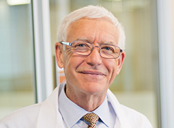
A Conversation About Pediatric Cancer Research and Treatment

$
0
0

Each year, more than 10,000 children in the U.S. are diagnosed with cancer, a figure that has slowly but steadily risen over the past several decades. Today, thanks to improved care, more kids are beating cancer than ever before. Still, more research is desperately needed to root out the causes of pediatric cancers, which differ greatly from adult cancers, and to leverage these findings into new, innovative treatments to help more kids lead longer, healthier lives.

Van Andel Institute (VAI) is honored to hold A Conversation about Pediatric Cancer hosted by Carol Van Andel, an educational and philanthropic event that will spotlight the exciting work underway in Grand Rapids to fight back against these devastating diseases. Guests will hear from the people on the front lines of these efforts, including Dr. Matt Steensma, who conducts research at the Institute and treats patients at Spectrum Health Helen DeVos Children’s Hospital, and Dr. Jenna Gedminas, a
clinical pediatric hematology/oncology fellow at Helen DeVos Children’s Hospital and research fellow in the lab of Dr. Patrick Grohar at the Institute. Dr. James Fahner, Division Chief of Pediatric Hematology/Oncology at Helen DeVos Children’s Hospital, will give the keynote address.

Registration

Title Sponsor

Silver Sponsor

Copper Sponsor

The post A Conversation About Pediatric Cancer Research and Treatment appeared first on VAI.

Disease profile: Pancreatic cancer

$
0
0

Pancreatic cancer is an aggressive, tough-to-treat disease in which malignant cells develop in the pancreas, a small gland located near the stomach that is responsible for producing hormones and digestive fluids. It is the third deadliest cancer in the U.S., claiming more than 40,000 lives each year. By 2020, it is expected to surpass colorectal cancers as the second-leading cause of cancer-related death.

Because it rarely causes noticeable symptoms in its early stages, pancreatic cancer often isn’t diagnosed until it is quite advanced. By this point, cancer cells may have spread to other parts of the body — a process called metastasis — further complicating an already challenging treatment process.

Current treatments for pancreatic cancer are largely limited to chemotherapy and surgery, with effectiveness based in part on how widespread cancer cells are in the body (for more on treatment, please see the American Cancer Society’s pancreatic cancer resources here; all treatment decisions should be made in close consultation with an oncologist).

Risk factors

  • Smoking
  • Obesity
  • Family history of pancreatic cancer or pancreatitis (a condition in which the pancreas is inflamed)
  • Having diabetes or chronic pancreatitis
  • Certain hereditary conditions (see here for a list)

How can research help?

Designing better tests: Right now, diagnosing pancreatic cancer early on is difficult at best and impossible at worst. Scientists like Van Andel Research Institute’s Dr. Brian Haab are working to develop simple blood tests that look for chemical signatures left by pancreatic cancer, which would enable doctors to detect this deadly disease much earlier. The sooner cancer is detected, the sooner treatment can begin, giving patients a better chance at remission (read more about Dr. Haab’s work in this area here).

Differentiating between subtypes: Understanding the tiny molecular differences that distinguish one pancreatic cancer from another gives scientists an important foundation upon which to develop new treatments. The Cancer Genome Atlas (TCGA) is a notable example. In 2017, this National Cancer Institute-led, multi-institutional effort reported results from in-depth analysis of 150 pancreatic cancer tumors, which confirmed major molecular drivers of the disease while also identifying several new features that may one day lead to more precise, personalized therapies.

Translating discovery into treatment: No two pancreatic cancers are identical, just as no two people are identical. That’s why scientists and physicians are working to design treatments that are tailored to individual patients based on the molecular makeup of their tumors, a change that one day could result in better care with fewer side effects.

Resources

Pancreatic cancer treatment — National Cancer Institute

ClinicalTrials.gov

The post Disease profile: Pancreatic cancer appeared first on VAI.

That’s a wrap! Van Andel Research Institute featured in Stand Up To Cancer telecast

$
0
0

Last Friday was a day to remember, one that will affect lives for years to come.

We were thrilled to be a part of the 2018 Stand Up To Cancer (SU2C) telecast, which raised a record-breaking $123 million for cancer research.

The Institute has worked closely with Stand Up To Cancer since 2014, when it became home to the Van Andel Research Institute–Stand Up To Cancer Epigenetics Dream Team, a multi-institutional effort to move more effective cancer therapies into clinical trials and onto the patients who need them most (read more about the team and its history here).

If you watched the telecast, you saw our own Ann Schoen, a VAI employee for 22 years, who was featured in the biennial special. She was part of the Everyday Heroes segment honoring cancer survivors.

For those who missed the telecast, we have you covered. You can watch it below, along with behind-the-scenes footage of Ann’s story and the digital show that preceded the broadcast.

Learn more about the VARI–SU2C Epigenetics Dream Team here.

The post That’s a wrap! Van Andel Research Institute featured in Stand Up To Cancer telecast appeared first on VAI.

Large-scale cancer study locates “light switches” in the “dark” regions of the genome

$
0
0
Many of cancer’s long-buried secrets are now out in the open thanks to a collaborative effort to catalog the genetic switches that govern life

The instructions for initiating and maintaining life are contained in DNA, a 6-foot-long, ladder-like molecule that determines every facet of our being from hair color to inherent risk for disease.

Yet only 2 percent of this DNA contains instructions for building proteins, the molecular workforce responsible for carrying out the directives written in our genes. The remaining bulk of our genetic material is “dark matter,” which for a long time had no defined function.

That all changed in 2012 with a series of discoveries from the ENCODE project that revealed these dark, or non-coding, regions are home to an entire system of molecular “light switches” that exert control over the genes that make us who we are and govern the behavior of cells.

Now, a collaborative team led by Stanford University’s Dr. Howard Y. Chang and Dr. William J. Greenleaf and including Van Andel Research Institute’s Dr. Peter W. Laird has developed the first comprehensive map detailing the accessibility of these switches and their role in cancer development. The study was part of The Cancer Genome Atlas (TCGA), a collaborative, National Institutes of Health-led effort to molecularly map cancer that wrapped up earlier this year.

“Cancers thrive on genetic discord, some of which occurs in these ‘dark’ regions,” Laird said. “This new resource will go a long way in helping us find new vulnerabilities in cancer and target them for treatment.”

The findings, published today in Science, build on ENCODE’s foundations by offering a detailed compendium of known and newly discovered switches and their locations on the genome, information that could greatly help scientists find better ways to combat cancer.

In the end, it all comes down to access. Each cell in the body contains a nearly exact copy of DNA, tightly wrapped and packaged in order to fit its entire length into the nucleus, the microscopic control center of the cell. To do this, DNA loops tightly around proteins called histones, curling tighter and tighter into a compact form called chromatin.

In order for genetic instructions to be carried out, specific areas of the chromatin must be accessible to special proteins called transcription factors, which flip the switches in non-coding regions to activate genes. In all, the team found hundreds of mutations that alter the control of gene activity in cancer. Some of these changes may make cancer tougher to treat but, thanks to today’s findings, they may also offer new therapeutic targets.

“Tiny variations in non-coding regions, often far away on the DNA strand from the gene they affect, play a massive role in every aspect of biology, including cancer development,” said Dr. Gerhard Coetzee, a VARI professor and expert on non-coding regions who was not involved in this study. “This map could be a game-changer, not only for improving our understanding of basic biology but more importantly, for patients who desperately need improved ways to fight back against cancer.”

Read more about this work here:

Study identifies link between DNA-protein binding, cancer onset—Stanford University

The post Large-scale cancer study locates “light switches” in the “dark” regions of the genome appeared first on VAI.

Viewing all 60 articles
Browse latest View live


Latest Images